Free-roaming Przewalski’s horses of Central Asia are often called the last of the wild horses, the only living equines never domesticated. But a new genetic analysis of ancient horse bones suggests that these horses have a tamed ancestor after all, making them feral rather than wild.
The findings also debunk the idea that these domesticated ancestors — known as Botai horses —gave rise to all other modern horses. That leaves the progenitors of today’s domesticated horses a mystery, researchers report online February 22 in Science.
The earliest known domesticated horses were those of the ancient Botai people in northern Kazakhstan (SN: 3/28/09, p. 15). Botai sites dating to around 5,500 years ago are scattered with remnants of harnesses and pots with horse-milk residue, suggesting the animals provided both transportation and food.
To see how Botai horses relate to today’s steeds, evolutionary geneticist Ludovic Orlando of the Natural History Museum of Denmark in Copenhagen and colleagues analyzed DNA from 88 horses spanning the last 5,000 years or so across Europe and Asia. Horses from the last 4,000 years had less than 3 percent Botai ancestry, suggesting that different and unknown horses founded today’s populations. But Botai horses are direct ancestors of Przewalski’s horses, the study found.
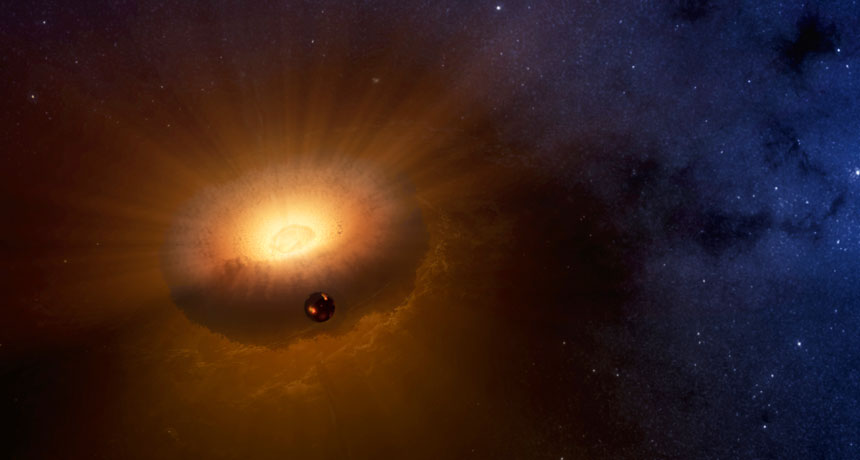
The moon might have formed from the filling during Earth’s jelly doughnut phase.
Around 4.5 billion years ago, something hit Earth, and the moon appeared shortly after. A new simulation of how the moon formed suggests it took shape in the midst of a hot cloud of rotating rock and vapor, which (in theory) forms when big planetary objects smash into each other at high speeds and energies. Planetary scientists Simon Lock of Harvard University and Sarah Stewart of the University of California, Davis proposed this doughnut-shaped planetary blob in 2017 and dubbed it a synestia (SN: 8/5/17, p. 5). Radiation at the surface of this swirling cloud of vaporized, mixed-together planet matter sent rocky rain inward toward bigger debris. The gooey seed of the moon grew from fragments in this hot, high-pressure environment, with a bit of iron solidifying into the lunar core. Some elements, such as potassium and sodium, remained aloft in vapor, accounting for their scarcity in moon rocks today.
After a few hundred years, the synestia shrank and cooled. Eventually, a nearly full-grown moon emerged from the cloud and condensed. While Earth ended up with most of the synestia material, the moon spent enough time in the doughnut filling to gain similar ingredients, Lock, Stewart and colleagues write February 28 in Journal of Geophysical Research: Planets . The simulation shakes up the prevailing explanation for the moon’s birth: A Mars-sized protoplanet called Theia collided with Earth, and the moon formed from distinct rubble pieces. If that’s true, moon rocks should have very different chemical compositions than Earth’s. But they don’t.
Other recent studies have wrestled with why rocks from the moon and Earth are so alike (SN: 4/15/17, p. 18). Having a synestia in the mix shifts the focus from the nature of the collision to what happened in its aftermath, potentially resolving the conundrum.
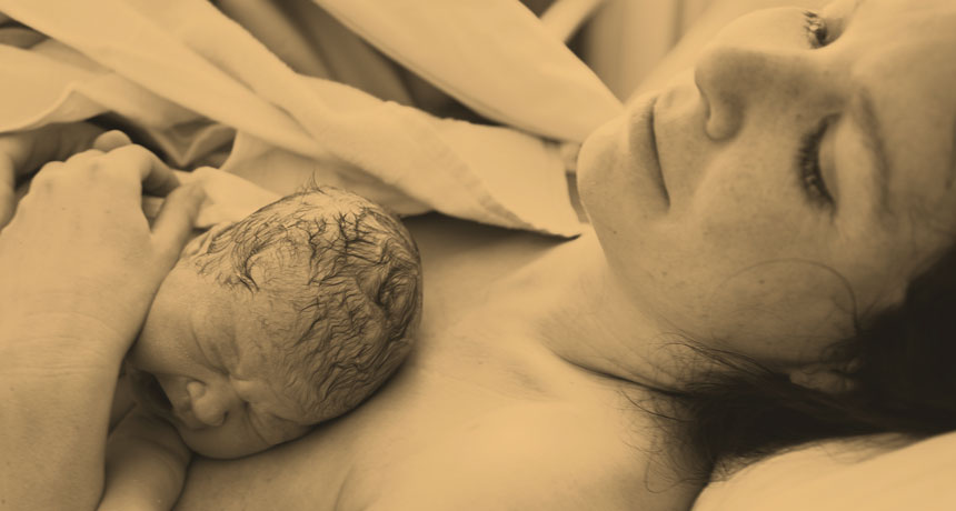
On the hormonal roller coaster of life, the ups and downs of childbirth are the Tower of Power. For nine long months, a woman’s body and brain absorb a slow upwelling of hormones, notably progesterone and estrogen. The ovaries and placenta produce these two chemicals in a gradual but relentless rise to support the developing fetus.
With the birth of a baby, and the immediate expulsion of the placenta, hormone levels plummet. No other physiological change comes close to this kind of free fall in both speed and intensity. For most women, the brain and body make a smooth landing, but more than 1 in 10 women in the United States may have trouble coping with the sudden crash. Those new mothers are left feeling depressed, isolated or anxious at a time society expects them to be deliriously happy. This has always been so. Mental struggles following childbirth have been recognized for as long as doctors have documented the experience of pregnancy. Hippocrates described a woman’s restlessness and insomnia after giving birth. In the 19th century, some doctors declared that mothers were suffering from “insanity of pregnancy” or “insanity of lactation.” Women were sent to mental hospitals.
Modern medicine recognizes psychiatric suffering in new mothers as an illness like any other, but the condition, known as postpartum depression, still bears stigma. Both depression and anxiety are thought to be woefully underdiagnosed in new mothers, given that many women are afraid to admit that a new baby is anything less than a bundle of joy. It’s not the feeling they expected when they were expecting.
Treatment — when offered — most commonly involves some combination of antidepression medication, hormone therapy, counseling and exercise. Still, a significant number of mothers find these options wanting. Untreated, postpartum depression can last for years, interfering with a mother’s ability to connect with and care for her baby.
Although postpartum depression entered official medical literature in the 1950s, decades have passed with few new options and little research. Even as brain imaging has become a common tool for looking at the innermost workings of the mind, its use to study postpartum depression has been sparse. A 2017 review in Trends in Neurosciences found only 17 human brain imaging studies of postpartum depression completed through 2016. For comparison, more than four times as many have been conducted on a problem called “internet gaming disorder” — an unofficial diagnosis acknowledged only five years ago. Now, however, more researchers are turning their attention to this long-neglected women’s health issue, peering into the brains of women to search for the root causes of the depression. At the same time, animal studies exploring the biochemistry of the postpartum brain are uncovering changes in neural circuitry and areas in need of repair.
And for the first time, researchers are testing an experimental drug designed specifically for postpartum depression. Early results have surprised even the scientists.
Women’s health experts hope that these recent developments signal a new era of research to help new moms who are hurting.
“I get this question all the time: Isn’t it just depression during the postpartum period? My answer is no,” says neuroscientist Benedetta Leuner of Ohio State University. “It’s occurring in the context of dramatic hormonal changes, and that has to be impacting the brain in a unique way. It occurs when you have an infant to care for. There’s no other time in a woman’s life when the stakes are quite as high.”
Brain drain Even though progesterone and estrogen changes create hormonal whiplash, pregnancy wouldn’t be possible without them. Progesterone, largely coming from the ovaries, helps orchestrate a woman’s monthly menstrual cycle. The hormone’s primary job is to help thicken the lining of the uterus so it will warmly welcome a fertilized egg. In months when conception doesn’t happen, progesterone levels fall and the uterine lining disintegrates. If a woman becomes pregnant, the fertilized egg implants in the uterine wall and progesterone production is eventually taken over by the placenta, which acts like an extra endocrine organ.
Like progesterone, estrogen is a normal part of the menstrual cycle that kicks into overdrive after conception. In addition to its usual duties in the female body, estrogen helps encourage the growth of the uterus and fetal development, particularly the formation of the hormone-producing endocrine system.
These surges in estrogen and progesterone, along with other physiological changes, are meant to support the fetus. But the hormones, or chemicals made from them, cross into the mother’s brain, which must constantly adapt. When it doesn’t, signs of trouble can appear even before childbirth, although they are often missed. Despite the name “postpartum,” about half of women who become ill are silently distressed in the later months of pregnancy.
Decades ago, controversy churned over whether postpartum depression was a consequence of fluctuating hormones alone or something else, says neuroscientist Joseph Lonstein of Michigan State University in East Lansing. He studies the neurochemistry of maternal caregiving and postpartum anxiety. Lonstein says many early studies measured hormone levels in women’s blood and tried to determine whether natural fluctuations were associated with the risk of postpartum depression. Those studies found “no clear correlations with [women’s] hormones and their susceptibility to symptoms,” he says. “While the hormone changes are certainly thought to be involved, not all women are equally susceptible. The question then became, what is it about their brains that makes particular women more susceptible?” Seeking answers, researchers have examined rodent brains and placed women into brain scanners to measure the women’s responses to pictures or videos of babies smiling, babbling or crying. Though hormones likely underlie the condition, many investigations have led to the amygdalae. These two, almond-shaped clumps of nerve cells deep in the brain are sometimes referred to as the emotional thermostat for their role in the processing of emotions, particularly fear.
The amygdalae are entangled with many structures that help make mothers feel like mothering, says neuroscientist Alison Fleming of the University of Toronto Mississauga. The amygdalae connect to the striatum, which is involved in experiencing reward, and to the hippocampus, a key player in memory and the body’s stress response. And more: They are wired to the hypothalamus, the interface between the brain and the endocrine system (when you are afraid, the endocrine system produces adrenaline and other chemicals that get your heart racing and palms sweating). The amygdalae are also connected to the prefrontal cortex and insula, involved in decision making, motivation and other functions intertwined with maternal instinct.
Fleming and colleagues have recently moved from studies in postpartum rodents to human mothers. In one investigation, reported in 2012 in Social Neuroscience, women were asked to look at pictures of smiling infants while in a functional MRI, which images brain activity. In mothers who were not depressed, the researchers found a higher amygdala response, more positive feelings and lower stress when women saw their own babies compared with unfamiliar infants.
But an unexpected pattern emerged in mothers with postpartum depression, as the researchers reported in 2016 in Social Neuroscience. While both depressed and not-depressed mothers showed elevated amygdala activity when viewing their own babies, the depressed mothers also showed heightened responses to happy, unknown babies, suggesting reactions to the women’s own children were blunted and not unique. This finding may mean that depressed women had less inclination to emotionally attach to their babies.
Mothers with postpartum depression also showed weaker connectivity between the amygdalae and the insula. Mothers with weaker connectivity in this area had greater symptoms of depression and anxiety. Women with stronger connectivity were more responsive to their newborns.
While there’s still no way to definitely know that the amygdalae are responding to postpartum chemical changes, “it’s very likely,” Lonstein says, pointing out that the amygdalae are influenced by the body’s reaction to hormones in other emotional settings.
Maternal rewards While important, the amygdalae are just part of the puzzle that seems to underlie postpartum depression. Among others is the nucleus accumbens, famous for its role in the brain’s reward system and in addiction, largely driven by the yin and yang of the neurotransmitters dopamine and serotonin. In studies, mothers who watched films of their infants (as opposed to watching unknown infants) experienced increased production of feel-good dopamine. The women also had a strengthening of the connection between the nucleus accumbens, the amygdalae and other structures, researchers from Harvard Medical School and their collaborators reported in February 2017 in Proceedings of the National Academy of Sciences.
That’s not entirely surprising given that rodent mothers find interacting with their newborn pups as neurologically rewarding as addictive drugs, says Ohio State’s Leuner. Rodent mothers that are separated from their offspring “will press a bar 100 times an hour to get to a pup. They will step across electrified grids to get to their pups. They’ve even been shown in some studies to choose the pups over cocaine.” Mothers find their offspring “highly, highly rewarding,” she says.
When there are postpartum glitches in the brain’s reward system, women may find their babies less satisfying, which could increase the risk for impaired mothering. Writing in 2014 in the European Journal of Neuroscience, Leuner and colleagues reported that in rats with symptoms of postpartum depression (induced by stress during pregnancy, a major risk factor for postpartum depression in women), nerve cells in the nucleus accumbens atrophied and showed fewer protrusions called dendritic spines — suggesting weaker connections to surrounding nerve cells compared with healthy rats. This is in contrast to other forms of depression, which show an increase in dendritic spines. Unpublished follow-up experiments conducted by Leuner’s team also point to a role for oxytocin, a hormone that spikes with the birth of a baby as estrogen and progesterone fall. Sometimes called the “cuddle chemical,” oxytocin is known for its role in maternal bonding (SN Online: 4/16/15). Leuner hypothesizes that maternal depression is associated with deficits in oxytocin receptors that enable the hormone to have its effects as part of the brain’s reward system.
If correct, the idea may help explain why oxytocin treatment failed women in some studies of postpartum depression. The hormone may simply not have the same potency in some women whose brains are short on receptors the chemical can latch on to. The next step is to test whether reversing the oxytocin receptor deficits in rodents’ brains relieves symptoms.
Leuner and other scientists emphasize that the oxytocin story is complex. In 2017, in a study reported in Depression & Anxiety, women without a history of depression who received oxytocin — which is often given to promote contractions or stem bleeding after delivery — had a 32 percent higher likelihood of developing postpartum depression than women who did not receive the hormone. In more than 46,000 births, 5 percent of women who did not receive the hormone were diagnosed with depression, compared with 7 percent who did.
“This was the opposite of what we predicted,” says Kristina Deligiannidis, a neuroscientist and perinatal psychiatrist at the Feinstein Institute for Medical Research in Manhasset, N.Y. After all, oxytocin is supposed to enhance brain circuits involved in mothering. “We had a whole group of statisticians reanalyze the data because we didn’t believe it,” she says. While the explanation is unknown, one theory is that perhaps the women who needed synthetic oxytocin during labor weren’t making enough on their own — and that could be why they are more prone to depression after childbirth.
But postpartum depression can’t be pinned to any single substance or brain malfunction — it doesn’t reside in one tidy nest of brain cells, or any one chemical process gone haywire. Maternal behavior is based on complex neurological circuitry. “Multiple parts of the brain are involved in any single function,” Deligiannidis says. “Just to have this conversation, I’m activating several different parts of my brain.” When any kind of depression occurs, she says, multiple regions of the brain are suffering from a communication breakdown.
Looking further, Deligiannidis has also examined the role of certain steroids synthesized from progesterone and other hormones and known to affect maternal brain circuitry. In a 2016 study in Psychoneuroendocrinology involving 32 new mothers at risk for postpartum depression and 24 healthy mothers, Deligiannidis and colleagues reported that concentrations of some steroids that affect the brain, also called neurosteroids, were higher in women at risk for developing depression (because of their past history or symptoms), compared with women who were not. The higher levels suggest a system out of balance — the brain is making too much of one neurosteroid and not enough of another, called allopregnanolone, which is thought to protect against postpartum depression and is being tested as a treatment. Treating pregnancy withdrawal Beyond mom
CASEZY IDEA/SHUTTERSTOCK Postpartum depression doesn’t weigh down just mom. Research suggests it might have negative effects on her offspring that can last for years. Risks include:
Newborns Higher levels of cortisol and other stress hormones More time fussing and crying More “indeterminate sleep,” hovering between deep and active sleep Infants and children Increased risk of developmental problems Slower growth Lower cognitive function Elevated cortisol levels Adolescents Higher risk of depression Tufts University neuroscientist Jamie Maguire, based in Boston, got interested in neurosteroids during her postgraduate studies in the lab of Istvan Mody at UCLA. Maguire and Mody reported in 2008 in Neuron that during pregnancy, the hippocampus has fewer receptors for neurosteroids, presumably to protect the brain from the massive levels of progesterone and estrogen circulating at that time. When progesterone drops after birth, the receptors repopulate.
But in mice genetically engineered to lack those receptors, something else happened: The animals were less interested in tending to their offspring, failing to make nests for them.
“We started investigating. Why are these animals having these abnormal postpartum behaviors?” Maguire recalls. Was an inability to recover these receptors making some women susceptible? Interestingly, similar receptors are responsible for the mood-altering and addictive effects of some antianxiety drugs, suggesting that the sudden progesterone drop after childbirth could be leaving some women with a kind of withdrawal effect.
Further experiments demonstrated that giving the mice a progesterone-derived neurosteroid — producing levels close to what the mice had in pregnancy — alleviated the symptoms.
Today, Maguire is on the scientific advisory board of Boston area–based Sage Therapeutics, which is testing a formulation of allopregnanolone called brexanolone. Results of an early clinical trial published last July in The Lancet assessed whether brexanolone would alleviate postpartum symptoms in women with severe postpartum depression. The study involved 21 women randomly assigned to receive a 60-hour infusion of the drug or a placebo within six months after delivery.
At the end of treatment, the women who received the drug reported a 21-point reduction on a standard scale of depression symptoms, compared with about 9 points for the women on a placebo. “These women got better in about a day,” says Deligiannidis, who is on the study’s research team. “The results were astonishing.”
In November, Sage Therapeutics announced the results of two larger studies, although neither has been published. Combined, the trials involved 226 women with severe or moderate postpartum depression. Both groups showed similar improvements that lasted for the month the women were followed. The company has announced plans to request approval from the U.S. Food and Drug Administration to market brexanolone in the United States. This is an important first step, researchers say, toward better treatments.
“We are just touching on one small piece of a bigger puzzle,” says Jodi Pawluski, a neuroscientist at the Université de Rennes 1 in France who coauthored the 2017 review in Trends in Neurosciences. She was surprised at the dearth of research, given how common postpartum depression is. “This is not the end, it’s the beginning.”
Ask a classroom of children to draw a scientist, and you’ll see plenty of Crayola-colored lab coats, goggles and bubbling beakers. That image hasn’t changed much since the 1960s. But the person wearing the lab coat is shifting.
A new analysis finds that more female scientists have appeared in kids’ drawings in recent decades — going from nearly nonexistent in the 1960s to about a third in 2016.
“A lot has changed since the 1960s,” says David Miller, a Ph.D. candidate in psychology at Northwestern University who reports the findings with colleagues March 20 in Child Development. The first of many “draw-a-scientist” studies asked nearly 5,000 children to draw a scientist between 1966 and 1977. “Of those 5,000 drawings,” Miller says, “only 28 … depicted a female scientist.” That’s just 0.6 percent.
Today, “more women are becoming scientists, and there’s some evidence that female scientists are being represented more in the media,” he says. For instance, in a content analysis of the magazine Highlights for Children, 13 percent of people pictured in science feature stories of the 1960s were women or girls, compared with 44 percent in the 2000s. To look for changes in children’s perceptions over time, the researchers conducted a meta-analysis, combining data from 78 studies that included a total of more than 20,000 U.S. children in kindergarten through 12th grade.
On average, 28 percent of children drew female scientists in studies conducted from 1985 to 2016, the researchers found.
What hasn’t changed much: Kids pick up stereotypes by gender as they grow up. At age 6, girls in the more recent studies drew female scientists about 70 percent of the time. By age 16, 75 percent drew male scientists.
“This is a critical period in which kids are learning stereotypes,” Miller says. “It’s important that teachers and parents present diverse examples of both male and female scientists.”
Editors’ note: This story was corrected on March 21, 2018, to note that by age 16, girls drew only 25 percent of scientists as female.
Off the Kohala coast on the Big Island of Hawaii, Christine Gabriele spots whale 875. The familiar propeller scar on its left side and the shape of its dorsal fin are like a telltale fingerprint. Gabriele, a marine biologist with the Hawaii Marine Mammal Consortium, confirms the whale’s identity against her extensive photo catalog. Both Gabriele and this male humpback have migrated to this Pacific Island from Southeastern Alaska.
In those Alaska summer feeding grounds, Gabriele sees the same 300 or so whales “again and again.” But winter brings more than 10,000 whales to the waters of Hawaii from all over the North Pacific. Spotting 875 is like finding a needle in a haystack. Gabriele is here today to focus on the slew of worrisome bumps on the familiar traveler’s flank. The bumps are separate from the usual ones bulging from the head of a humpback ( Megaptera novaeangliae ). Those iconic oversize hair follicles are thought to be part of the sensory system. The smaller body bumps look more like bad acne or an allergic reaction. Noted on rare occasions in the 1970s, the condition called nodular dermatitis has become much more prevalent. These days, Gabriele and colleagues see these skin lesions on over 75 percent of Hawaii’s humpback visitors. The bumps coincide with other suggestions of declining health in the whales. In the nearly three decades that Gabriele has been studying whales, she would not describe the animals as skinny. Now, often “you can see their shoulder blades,” she says. “They look angular rather than round.” Gabriele’s team is trying to figure out the cause of the bumps, comparing tissue samples from bumpy and nonbumpy whales. Several times per week, a small team sets out on the water, research permits in hand. Once a whale pod is spotted, Gabriele’s colleague Suzanne Yin zooms in with a camera and volunteer Kim New enlarges the image on her iPad, examining skin on the whale’s flanks and behind the blowhole to confirm if it’s bumpy or not. Gabriele carefully steers the boat so that Yin can shoot a biopsy dart from a crossbow. The dart “takes a little plug of skin and blubber … about the size of a pencil eraser,” Gabriele says. The dart bounces off the whale and floats until the researchers can grab it. When darted, some whales dive; others show no reaction at all.
Collaborators from the National Institute of Standards and Technology’s Hollings Marine Laboratory in Charleston, S.C., are analyzing the skin for trace elements. National Marine Fisheries Service lab staff are studying the blubber for organic pollutants like PCBs and flame retardants. Preliminary results suggest that bumpy whales differ from nonbumpy in levels of manganese and a few other trace elements. Gabriele eagerly awaits the full analyses to make sense of what she’s seeing among the migratory creatures.
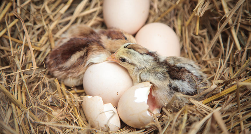
A chicken eggshell has a tricky job: It must protect a developing chick, but then ultimately let the chick break free. The secret to its success lies in its complex nanostructure — and how that structure changes as the egg incubates.
Chicken eggshells are about 95 percent calcium carbonate by mass. But they also contain hundreds of different kinds of proteins that influence how that calcium carbonate crystalizes. The interaction between the mineral crystals and the proteins yields an eggshell that’s initially crack-resistant, while making nanoscale adjustments over time that ultimately let a chick peck its way out, researchers report online March 30 in Science Advances. Researchers used a beam of ions to cut thin cross sections in chicken eggshells. They then analyzed the shells with electron microscopy and other high-resolution imaging techniques. The team found that proteins disrupt the crystallization of calcium carbonate, so that what seems at low resolution to be neatly aligned crystals is actually a more fragmented jumble. This misalignment can make materials more resilient: Instead of spreading unimpeded, a crack must zig and zag through scrambled crystals. Lab tests back up that finding: The researchers added a key shell-building protein called osteopontin to calcium carbonate to yield crystals like those seen in the eggshells. The presence of that protein makes calcium carbonate crystals form in a nanostructured pattern, rather than smooth and even crystal, study coauthor Marc McKee, a biomineralization researcher at McGill University in Montreal, and colleagues found.
The team also found structural variation on a minute scale throughout the eggshell, though it’s only about a third of a millimeter thick. Inner layers have less osteopontin, leading to bigger nanostructures. That may make the inner shell less resilient than the outer shell, which makes sense, McKee says. The outer shell needs to be hard enough to protect the chick, while the inner shell nourishes the developing chick.
Over time, the inner layers of the shell dissolve through a chemical reaction, releasing calcium to build a chick’s developing bones. The eggshell undergoes structural changes to facilitate that process, McKee and his colleagues found.
The researchers compared fertilized eggs incubated for 15 days to nonfertilized eggs. Over time, the nanostructures toward the inner shell became smaller in fertilized eggs, but remained the same in the nonfertilized eggs. The change gives the inside of the eggshell a bumpier texture, and by extension, more surface area. That provides more space for that shell-dissolving chemical reaction to take place, the researchers propose. The reaction also thins the shell overall, making it easier for a chick to break through from the inside when it’s time to hatch.
Advances in imaging technology are helping scientists find new details like this even in objects as familiar as a chicken eggshell, says Lara Estroff, a materials scientist at Cornell University who wasn’t part of the research. In connecting the eggshell’s functionality with its fine-grain structure, the new study could provide inspiration for designing new kinds of materials with specific properties.
Your brain might make new nerve cells well into old age.
Healthy people in their 70s have just as many young nerve cells, or neurons, in a memory-related part of the brain as do teenagers and young adults, researchers report in the April 5 Cell Stem Cell. The discovery suggests that the hippocampus keeps generating new neurons throughout a person’s life.
The finding contradicts a study published in March, which suggested that neurogenesis in the hippocampus stops in childhood (SN Online: 3/8/18). But the new research fits with a larger pile of evidence showing that adult human brains can, to some extent, make new neurons. While those studies indicate that the process tapers off over time, the new study proposes almost no decline at all. Understanding how healthy brains change over time is important for researchers untangling the ways that conditions like depression, stress and memory loss affect older brains.
When it comes to studying neurogenesis in humans, “the devil is in the details,” says Jonas Frisén, a neuroscientist at the Karolinska Institute in Stockholm who was not involved in the new research. Small differences in methodology — such as the way brains are preserved or how neurons are counted — can have a big impact on the results, which could explain the conflicting findings. The new paper “is the most rigorous study yet,” he says.
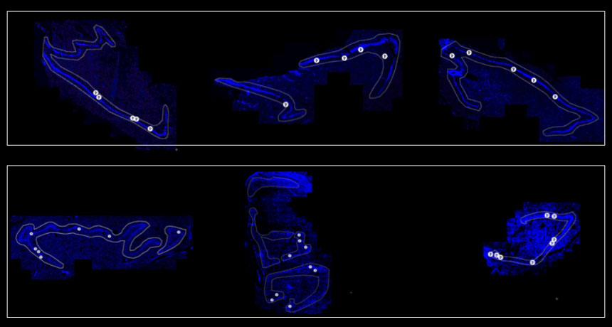
Researchers studied hippocampi from the autopsied brains of 17 men and 11 women ranging in age from 14 to 79. In contrast to past studies that have often relied on donations from patients without a detailed medical history, the researchers knew that none of the donors had a history of psychiatric illness or chronic illness. And none of the brains tested positive for drugs or alcohol, says Maura Boldrini, a psychiatrist at Columbia University. Boldrini and her colleagues also had access to whole hippocampi, rather than just a few slices, allowing the team to make more accurate estimates of the number of neurons, she says. To look for signs of neurogenesis, the researchers hunted for specific proteins produced by neurons at particular stages of development. Proteins such as GFAP and SOX2, for example, are made in abundance by stem cells that eventually turn into neurons, while newborn neurons make more of proteins such as Ki-67. In all of the brains, the researchers found evidence of newborn neurons in the dentate gyrus, the part of the hippocampus where neurons are born.
Although the number of neural stem cells was a bit lower in people in their 70s compared with people in their 20s, the older brains still had thousands of these cells. The number of young neurons in intermediate to advanced stages of development was the same across people of all ages.
Still, the healthy older brains did show some signs of decline. Researchers found less evidence for the formation of new blood vessels and fewer protein markers that signal neuroplasticity, or the brain’s ability to make new connections between neurons. But it’s too soon to say what these findings mean for brain function, Boldrini says. Studies on autopsied brains can look at structure but not activity.
Not all neuroscientists are convinced by the findings. “We don’t think that what they are identifying as young neurons actually are,” says Arturo Alvarez-Buylla of the University of California, San Francisco, who coauthored the recent paper that found no signs of neurogenesis in adult brains. In his study, some of the cells his team initially flagged as young neurons turned out to be mature cells upon further investigation.
But others say the new findings are sound. “They use very sophisticated methodology,” Frisén says, and control for factors that Alvarez-Buylla’s study didn’t, such as the type of preservative used on the brains.
Composting waste is heralded as being good for the environment. But it turns out that compost collected from homes and grocery stores is a previously unknown source of microplastic pollution, a new study April 4 in Science Advances reports.
This plastic gets spread over fields, where it may be eaten by worms and enter the food web, make its way into waterways or perhaps break down further and become airborne, says Christian Laforsch, an ecologist at the University of Bayreuth in Germany. Once the plastic is spread across fields, “we don’t know its fate,” he says. That fate and the effects of plastic pollution on land and in freshwater has received little research attention compared with marine plastic pollution, says ecologist Chelsea Rochman of the University of Toronto. Ocean microplastics have gained notoriety thanks in part to coverage of the floating hulk of debris called the great Pacific garbage patch (SN Online: 3/22/18).
But current evidence suggests that plastic pollution is as prevalent in land and freshwater ecosystems as it is in the oceans, where it’s found “from the equator to the poles,” says Rochman, author of a separate commentary on the state of plastic pollution research published in the April 6 Science. Plastic “is seen in the high Arctic, where we suspect it comes down in rain. We know it’s in drinking water, in our seafood and spread on our agricultural fields,” she says.
Laforsch and his colleagues looked at several different kinds of biowaste that’s composted and spread on farmland in Germany, including household compost and grass clippings, supermarket waste and grain silage leftover from biogas production.
Compost samples taken from supermarket waste contained the greatest amount of plastic particles, with 895 pieces larger than 1 millimeter found per kilogram of dry weight. Household compost in contrast contained 20 and 24 particles per kg of dry weight, depending on the size of the sieves used to sift the compost. The detritus included bits of polyester and a lot of styrene-based polymers, commonly used in food packaging. Almost no particles were found in samples of silage from biogas production. “I never thought about plastic in compost ending up as fertilizer. But when you think about it, it makes sense,” says environmental scientist Ad Ragas of Radboud University in Nijmegen, The Netherlands, who wasn’t involved in the work. A crate of rotting cucumbers wrapped in plastic that gets chucked, those stickers on every tomato in a bunch — that packaging doesn’t disappear.
Ragas says compost probably doesn’t contribute as much plastic to the environment as other sources, such as sewage treatment plant sludge, which contains polyester debris from clothes washers, and runoff from streets, which can be loaded with particles of synthetic rubber used in tires. But the compost contribution deserves investigation, Ragas says. “This triggers a lot of questions we haven’t studied yet.”
Those questions include possible effects on different organisms, from plants to earthworms to birds to people, Rochman says. Those effects will likely differ depending on the kind of plastic, which varies depending on its starting polymer and the additives used to impart certain qualities such as flexibility, sturdiness or durability under ultraviolet light.
“We are not saying we should get rid of all plastics,” Rochman says. “But we do need to start thinking of plastics as a persistent global pollutant.”
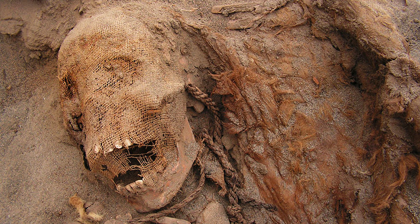
A hellishly unprecedented scene — what anthropologists suspect is the largest known child sacrifice — has been unearthed on a bluff overlooking Peru’s northern shoreline.
Around 550 years ago, members of the Chimú empire ritually killed and buried at least 140 children, ages 5 to 14, and 200 young llamas, says a team led by Gabriel Prieto of the National University of Trujillo in Peru and John Verano of Tulane University in New Orleans.
“There are no other examples of child sacrifices anywhere in the world that compare to the magnitude of this Chimú event,” Verano says. The discovery was announced April 26 by National Geographic in Washington, D.C. Except for a few incomplete skeletons, excavated children and llamas displayed cuts on their breast bones and dislocated ribs indicating that their chests had been sliced open. Three adults buried nearby on the bluff, including two women with violent head wounds, may have participated in the sacrifice.
Radiocarbon dating, mainly of ropes left around the llamas’ necks, puts the event at around 1450, shortly before the Inca conquered the Chimú in 1470.
A dried mud layer covering some of the sandy graves possibly resulted from flooding caused by massive rains. Agricultural crises triggered by repeated flooding might have led Chimú leaders to sacrifice children to their gods, Verano suggests.
For some neutron stars, the quickest way to cool off isn’t with a frosty beverage, but with lightweight, subatomic particles called neutrinos.
Scientists have spotted the first solid evidence that some neutron stars, the collapsed remnants of exploded stars, can rapidly cool their cores by emitting neutrinos. The result adds to evidence that scientists are gathering to understand the ultradense matter that is squished deep within a neutron star’s center.
The new evidence comes from a neutron star that repeatedly gobbled material from a neighboring star. The neutron star rapidly cooled off after its meals, scientists determined. X-rays emitted by the neutron star showed that the fast cooldown rate was consistent with a theorized effect called the direct Urca process, in which neutrinos quickly ferry energy away from a collapsed star, astrophysicist Edward Brown and colleagues report in the May 4 Physical Review Letters. Neutron stars are known to emit neutrinos by a similar process that cools the star slowly. But previously, there wasn’t clear evidence for faster cooling. The team analyzed observations of the neutron star, located about 35,000 light-years from Earth, as it cooled during a 15-year interlude between feeding sessions. Neutrinos carried away energy about 10 times faster than the rate energy is radiated by the sun’s light — or about 100 million times quicker than the slow process, says Brown, of Michigan State University in East Lansing.
Although some other neutron stars have shown hints of such a quick chill, “this is basically the first object for which we can see the star actively cooling before our eyes,” says astrophysicist James Lattimer of Stony Brook University in New York, who was not involved with the research.
The direct Urca process, named by physicists George Gamow and Mário Schenberg in the 1940s, took its moniker from the now-defunct Urca casino in Rio de Janeiro. “The joke being that this process removes heat from the star the way the casino removes money from tourists’ pockets,” Brown says. In the process, neutrons in the star’s core convert into protons and emit electrons and antineutrinos (the antimatter partners of neutrinos). Likewise, protons convert into neutrons and emit antielectrons and neutrinos. Because neutrinos and antineutrinos interact very rarely with matter, they can escape the core, taking energy with them. “The neutrino is a thief; it robs energy from the star,” says physicist Madappa Prakash of Ohio University in Athens, who was not involved with the research.
The observation may help scientists understand what goes on deep within neutron stars, the cores of which are squeezed to densities far beyond those achievable in laboratories. Although the simplest theory holds that the cores are crammed with neutrons and a smaller number of protons and electrons, scientists have also proposed that the collapsed stars may consist of weird states of matter, containing rare particles called hyperons or a sea of free-floating quarks, the particles that make up protons and neutrons (SN: 12/23/17, p. 7).
The direct Urca process can happen only if the fraction of protons in the center of the neutron star is larger than about 10 percent. So if the process happens, “that already tells us a lot,” says astrophysicist Wynn Ho of Haverford College in Pennsylvania, who was not involved in the research. Such observations could eliminate theories that would predict lower numbers of protons.
However, the scientists weren’t able to determine the mass of the neutron star, limiting the conclusions that can be drawn. But, says Prakash, if the mass of such a quickly cooling neutron star is measured, the neutron star’s interior makeup could be nailed down.